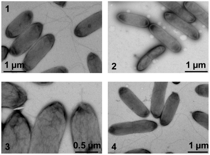
Figure 3. Transmission electron micrographs of S. oneidensis wild type and flagellar mutantstrains.
Cells were stained with 1% phophotungstic acid and applied to TEM. 1. The wild type. 2.ΔflgK, aflagellated. 3. ΔfliD, truncated filaments. 4. ΔSO3234, flagella indistinguishable from that of the wild type.

