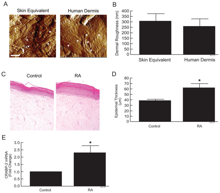
Figure 1.
Organization of collagen fibrils and RA responsiveness in skin equivalent cultures. (a) Skin equivalent cultures and human skin were embedded in OCT and cryo-sections (7 μm) were analyzed by atomic force microscopy (AFM). AFM images were obtained using a Multimode Nanoscope IIIa AFM (Veeco Instrument Inc., CA), as described in Experimental Procedures. Images are representative of three experiments. Bar = 1μm. Arrows indicate collagen bundles in skin equivalent cultures and human skin (b) Skin dermal roughness was determined by Nanoscope IIIa software. Data are expressed as mean±SEM, N=4. (c) Skin equivalent cultures were treated with all-trans retinoic acid (10μM) for 7 days. Cultures were embedded in OCT and cryo-sections (7 μm) were stained with H&E. Lines indicate dermal-epidermal boundary. Images are representative of three experiments. (d) Average vertical height of the stratified layers of keratinocytes was quantified by computerized image analysis. Data are expressed as mean±SEM, N=3, *p<0.05. (e) CRABP-2 mRNA levels were quantified by real-time RT-PCR and were normalized to mRNA for 36B4, a ribosomal protein used as an internal control for quantitation. Data are expressed as mean±SEM, N=3, *p<0.05.

