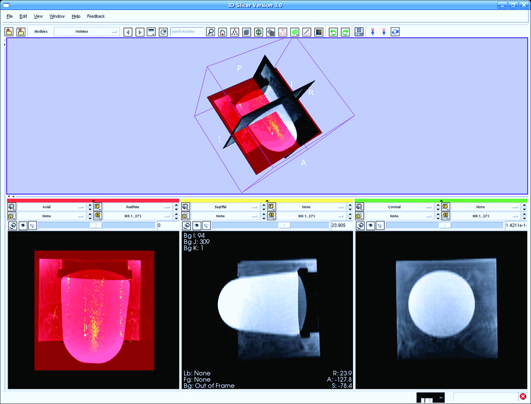
Fig. 8.
Screen snapshot of 3D-Slicer, the display software package. The bottom left image displays a snapshot during the real-time imaging, processing, and display. The FUS heating effect, which is updated in real-time, is shown in yellow superimposed on the baseline axial image of the gel. The transducer is located in the upper portion of the bottom left of the image.

