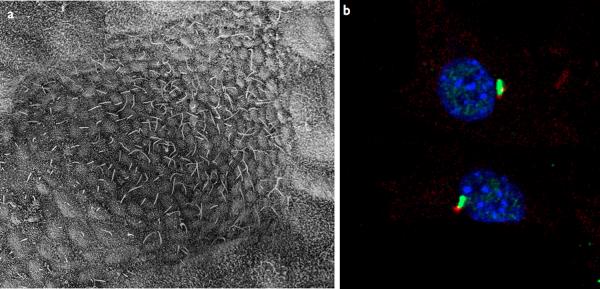
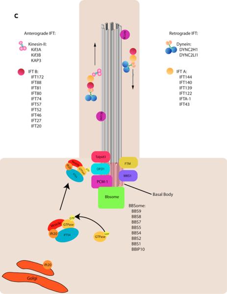
Figure 1. Cilia structure and IFT.
a–b | Examples of types of mammalian cilia. a | The long cilia of the node at E7.5 are required for left-right asymmetry. b | Primary cilia in embryonic fibroblasts in green with the basal body in red.
c | Cargo is transported from the base to the tip of the cilium along the microtubule axoneme by Kinesin-2 together with the IFT-A and IFT-B complexes. Dynein mediates the return of IFT cargo to the base of the cilium1, 14. IFT-B proteins IFT-20 and IFT-54 may also participate in the trafficking of membrane vesicles from the Golgi to the ciliary membrane together with small GTPases128. Other small GTPases, including Arl13b also localize to cilia. While its precise trafficking role is not known, Arl13b is required for axoneme structure129. Certain basal body proteins also influence ciliary trafficking. Among these are components of the Bbsome, named their association with Bardet-Biedl syndrome (BBS). The precise functions of BBS proteins in cilia formation are unclear as they are not individually required for primary cilia formation, however they may function to promote loading of cargo to the ciliary axoneme17. Other basal body associated protein such as MKS1, FTM, OFD1, and Talpid3 are required for cilia formation, though how they regulate ciliogenesis has not been defined (For a review, see10).


