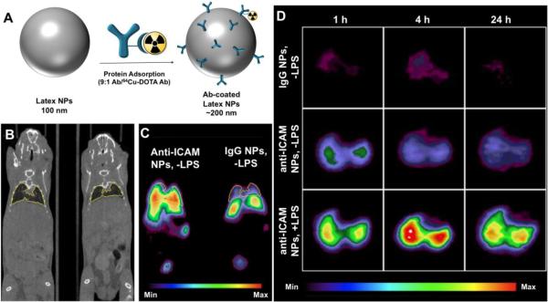
Figure 4.
(A) Preparation of 200 nm Ab-coated polymeric latex NPs for 64Cu PET imaging. 64Cu was chelated to 1,4,7,10-tetraazacyclododecane-1,4,7,10-tetraacetic acid (DOTA) preconjugated to IgG, and then mixed with unlabeled anti-ICAM or IgG Ab (1:9) for adsorption onto NP surface (~250 Ab/NP). (B) MicroCT (coronal slice) and (C) microPET (coronal slice) images of 2 naïve mice (−LPS) injected with ICAM-targeted NPs or control IgG-coated NPs 1 h p.i. (D) Representative decay-corrected transverse micro-PET images of naïve mice (−LPS) and LPS-challenged mice (+LPS) at 1, 4, and 24 h after NP administration. Uptake intensity map is normalized to the highest pixels in the LPS-challenged mice. Figure modified from [75]. © 2008 Society of Nuclear Medicine.

