Fig. 2.

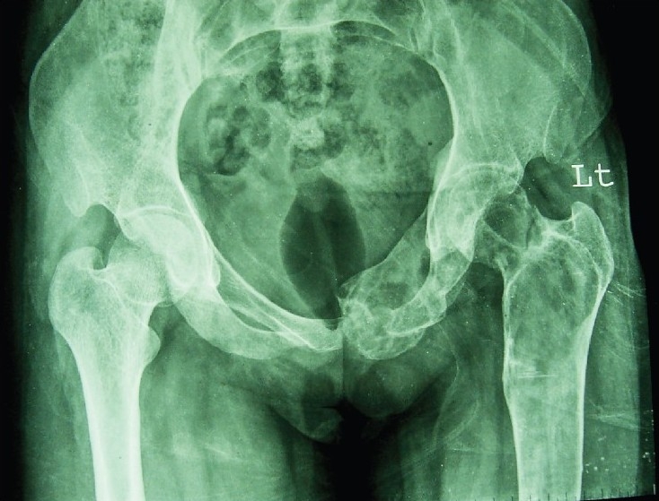
X-ray pelvis AP view showing multiple expansile lytic lesion with fish net appearance involving left ischium, pubis, left neck of femur and greater trochanter.

X-ray pelvis AP view showing multiple expansile lytic lesion with fish net appearance involving left ischium, pubis, left neck of femur and greater trochanter.