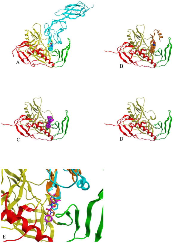
Figure 2.
The four constructs used in MD simulation are shown in panels A – D with the three gp120 domains; inner, outer and bridging sheet colored as red, yellow and green ribbons, respectively. A) The gp120-CD4 complex (GCD2) with CD4 shown in blue with Phe 43 depicted as space-filling model. B) The gp120-CD4M47 complex (GCD3) with the scorpion toxin derived mini-protein shown in orange with the residue 23 biphenyl depicted as space-filling model. C) NBD-556 docked to gp120 (GPO3_NBD) with NBD-556 shown as purple space filling model. D) the unbound gp120 structure (GPO3) E) Comparison of the NBD-556 (purple), biphenyl (orange) and Phe-43 (cyan) as bound in the cavity gp120 for GPO3_NBD, GCD3 and GCD2, respectively.

