Figure 5.
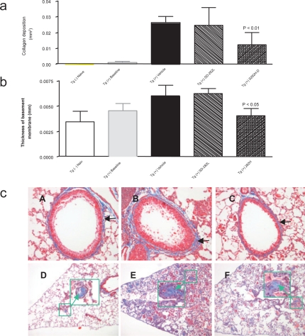
Effect of SD-282 on fibrosis induced in the CC10:IL-13 transgenic asthma model analyzed after Masson’s trichrome stain. [a] SD-282 at high-dose significantly reduces fibrosis of lung parenchyma compared with the vehicle-treated group (p < 0.01). The noted values represent the MEAN ± SD on a minimum of eight animals. [b] Thickness of basement membrane following Masson’s trichrome stain. SD-282 at high-dose significantly reduces subepithelial fibrosis compared with the vehicle-treated group (p < 0.05).The noted values represent the MEAN ± SD on a minimum of eight animals. [c] Masson’s trichrome stain, x200 showing lung sections from Tg (+) baseline (A), Tg (+) vehicle (B) and Tg (+) SD-282H (C). Very mild collagen deposition stained in blue (black arrow) around the basement is seen in Tg (+) mice in baseline (A). Enhanced accumulation of collagen stained in blue (black arrow) is seen in the vehicle-treated Tg (+) mice (B). Less collagen accumulation and reduced thickening in the basement membrane (black arrow) are seen in the SD-282 high-dose treated Tg (+) mice (C). Masson’s trichrome stain, x100 and x400 (in large green box with arrow) showing lung sections from Tg (+) baseline (D), Tg (+) vehicle (E), and Tg (+) SD-282H (F). Mild fibrotic change is seen in the Tg (+) mice in baseline (D). Numerous fibrotic beads with collagen deposition stained in blue (green box) and elongated fibroblasts are seen in the vehicle-treated Tg (+) mice (E). Reduced fibrotic beads are seen in SD-282 high-dose treated Tg (+) mice (F).

