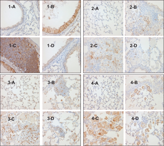
Figure 6.
Effect of p38 MAPK inhibition on immune cell infiltration and activation in CC10:IL-13 mice [1] p-p38 MAPK IHC stain, x400 showing no p-p38 MAPK activation in Tg (−) mice (1-A), enhanced p-p38 MAPK activation in the epithelial cells of airway (1-B) and infiltrated lymphocytes(1-C) in the vehicle-treated Tg (+) mice and completed inhibition of p-p38 MAPK activation in the epithelial cells in the SD-282 high-dose treated Tg (+) mice (1-D). [2] IL-1β IHC stain, x400 showing no IL-1β expression in Tg (−) mice (2-A), mild IL-1β expression in the PMN cells in Tg (+) mice in baseline (2-B), increased IL-1β expression in the vehicle-treated Tg (+) mice (2-C) and less IL-1β expression in the PMN cells in the SD-282 high-dose treated Tg (+) mice (2-D). [3] CD3 IHC stain, x400 showing few CD3+ T-lymphocytes in the lung of Tg (−) mice (3-A), mild CD3+ T-lymphocyte infiltration in the lung of Tg (+) mice in baseline (3-B), markedly increased number of CD3+ T-lymphocyte in the lung of Tg (+) mice treated with vehicle (3-C) and significantly decreased number of CD3+ T-lymphocytes in the lung of Tg (+) mice treated with the high-dose of SD-282 (3-D). [4] F4/80 IHC, x400 showing no activated macrophages in the lung of Tg (−) mice (4-A), few activated macrophages in the lung of Tg (+) mice in baseline (4-B), markedly increased number of activated macrophages in the lung of Tg (+) mice treated with vehicle (4-C), and decreased number of activated macrophages in the lung of Tg (+) mice treated with high-dose of SD-282 (4-D).

