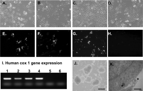
FIGURE 2.
A–D, phase contrast microscopy showing the morphology of 1.1B4, 1.1E7, 1.4E7, and parental PANC-1 cell lines grown as monolayers in culture. E, F, and G, immunohistochemistry showing positive staining for insulin in 1.1B4, 1.1E7, and 1.4E7 cell lines (E, F, and G, respectively). H, PANC-1 cells are negative for insulin. Slides were analyzed by fluorescence microscopy using an IX51 Olympus microscope (×20 magnification). I, RT-PCR showing expression of the human cox-1 gene in 1.1B4, 1.1E7, 1.4E7, and parental PANC-1 cell lines (lanes 1–4, respectively). Lane 5 contained no maize mosaic virus (MMV) reverse transcriptase; RNA from mouse pancreas was used as negative control (lane 6). J, phase contrast of 1.1B4 cells showing aggregation into the pseudoislet structure if maintained in suspension culture using ultralow attachment flat-bottomed 6-well plates (×10 magnification; scale bar, 200 μm) K, transmission electron micrographs showing the presence of dark insulin granules (asterisks) in the cytoplasm and external surface of 1.1B4 cells; scale bar, 2 μm.

