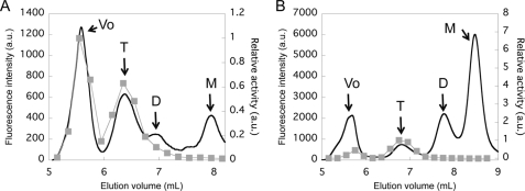
FIGURE 4.
Size exclusion chromatography of GAL-SNAP (A) and GUS-SNAP (B) synthesized in vitro at 37 °C for 2 h. The black lines show the fluorescence signal (left axis) and gray squares show the enzymatic activity of eluted fraction (right axis). Maximum activity was defined as 1. The elution volumes of void volume (Vo), tetramer (T), dimer (D), and monomer (M) estimated from the molecular weight standards (supplemental Fig. S4) are indicated.

