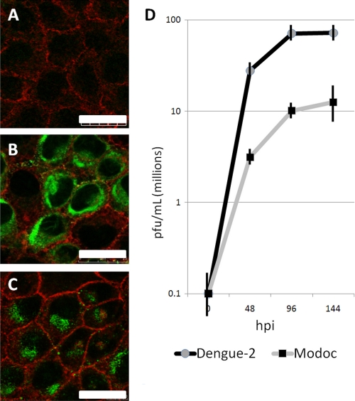
FIGURE 2.
Flaviviruses establish a productive infection in renal epithelial cells. Dengue-2 and Modoc virus-infected MDCK cells were fixed at 48 hpi and stained with phalloidin-TRITC (red, actin) and antibody Di-4G2-15 (green) flavivirus Envelope protein. A, mock − infected cells lack positive staining for virus protein. Massive flavivirus Envelope protein accumulation in cytoplasmic vesicles within the ER-rich perinuclear region of both Dengue-2 virus (B) and Modoc virus (C)- infected cells demonstrates the establishment of flavivirus translation and assembly. D, qRT-PCR of infected cell supernatant demonstrates rapid replication and release of virus.

