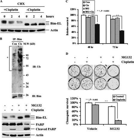
FIGURE 5.
Effects of cisplatin and proteasome inhibitors on Bim-EL degradation and cell death. A, Western blot analyses of Bim-EL expression. RMG-1 cells were treated with 2 μg/ml cycloheximide (CHX) for the indicated time periods in the presence and absence of 10 μm cisplatin for 48 h. Actin was used as a loading control. B, RMG-1 cells were left untreated or treated with 10 μm cisplatin (Cis) for 24 h (upper panel). Bim was immunoprecipitated, and Western blot analyses with anti-Bim and ubiquitin (Ub) antibodies were performed. The asterisk indicates ubiquitinated Bim appearing as a smear of bands with a higher molecular weight. RMG-1 cells were left untreated or treated with 10 μm cisplatin in the presence (+) or absence (−) of the proteasome inhibitor MG132 (250 nm) for 48 h (lower panel). Actin was used as a loading control (Con). IB, immunoblotting. C, MTT analyses of growth inhibition. RMG-1 cells were left untreated or treated with 10 μm cisplatin in the presence or absence of 250 nm MG132 for 48 or 72 h. D, colony formation analyses of cell growth. RMG-1 cells were left untreated or treated with 10 μm cisplatin in the presence or absence of 250 nm MG132 (MG) for 3 h. 400 cells/well were seeded in 6-well plates and grown for 12 days, followed by crystal violet staining. Quantification of survival colonies is shown (lower panel). The plating efficiencies of drug-treated wells were normalized to those of control wells. The plating efficiency of the control wells was arbitrarily established as 100%.

