FIGURE 2.
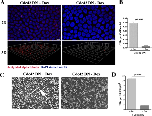
Dominant Negative Expression of Cdc42 Inhibits Ciliogenesis. A, MDCK cells expressing dominant negative (DN) Cdc42 in the presence and absence of doxycycline were grown on Transwell filters for 14 days. Using an antibody against acetylated α-tubulin (red), ciliogenesis was examined by confocal microscopy combined with three-dimensional (3D) reconstruction of the stacked series. Ciliogenesis was virtually completely inhibited when Cdc42 DN protein was expressed (in the absence of doxycycline (Dox)). DAPI (blue) is a nuclear stain. DAPI staining is at a different level in the cell than staining for acetylated α-tubulin but is included in the merged figure to delineate individual cells and allow for statistical analysis. Scale bar = 5 μm. B, quantification of ciliogenesis was performed using a ratio of cilia to cell nuclei. Significantly fewer cilia were seen in the MDCK cells expressing Cdc42 DN protein (-Dox). C, Cdc42 DN cells were grown on Transwell filters in the presence (+) and absence (-) of doxycycline for 14 days. The cells were fixed in glutaraldehyde, and SE microscopy was performed (Phillips XL20). Confirming the results in A, primary cilia were rarely seen when Cdc42 DN protein was expressed (-Dox). Scale bar = 1.0 μm. D, quantification of ciliogenesis was performed by counting the number of cilia per surface area, as individual cells could not be identified. Significantly fewer cilia were seen in the MDCK cells expressing Cdc42 DN protein (-Dox)..

