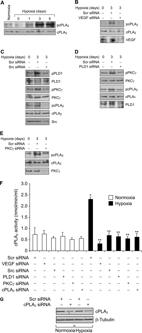
FIGURE 6.
Hypoxia stimulates cPLA2 phosphorylation via Src-PLD1-PKCγ signaling axis in retina. A, C57BL/6 mice pups were exposed to 75% oxygen from P7 to P12 at which time they were returned to normoxia to develop relative hypoxia for the indicated time periods; eyes were enucleated, retinas were isolated, and tissue extracts were prepared and analyzed by Western blotting for cPLA2 phosphorylation using its phosphospecific antibodies. The blot was reprobed with anti-cPLA2 antibodies for normalization. B, after exposure to hyperoxia, mice pups were returned to normoxia and administered 1 μg/0.5 μl scrambled (Scr) or VEGF siRNA at P12 and P13 by intravitreal injections. Retinas were isolated at P15, and extracts were prepared and analyzed by Western blotting for cPLA2 phosphorylation using its phosphospecific antibodies. The blot was reprobed sequentially with anti-cPLA2 and anti-VEGF antibodies for normalization or to show down-regulation of VEGF by its siRNA. C, all conditions were the same as in B except that mice pups were administered scrambled or Src siRNA at P12 and P13 by intravitreal injections, and at P15 the retinas were isolated, and extracts were prepared and analyzed for phosphorylation of PLD1, PKCγ, and cPLA2 using their phosphospecific antibodies. The blots were reprobed with anti-PLD1, anti-PKCγ, anti-cPLA2, or anti-Src antibodies either for normalization or to show the down-regulation of Src by its siRNA. D, all conditions were the same as in B except that mice pups were administered scrambled or PLD1 siRNA at P12 and P13 by intravitreal injections, and at P15 the retinas were isolated and extracts prepared and analyzed for phosphorylation of PKCγ and cPLA2 using their phosphospecific antibodies. The blots were reprobed with anti-PKCγ, anti-cPLA2, or anti-PLD1 antibodies either for normalization or to show the down-regulation of PLD1 by its siRNA. E, all conditions were the same as in B except that mice pups were administered scrambled or PKCγ siRNA at P12 and P13 by intravitreal injections, and at P15 the retinas were isolated and extracts prepared and analyzed for phosphorylation of cPLA2 using its phosphospecific antibodies. The blot was reprobed sequentially with anti-cPLA2 and anti-PKCγ antibodies either for normalization or to show the down-regulation of anti-PKCγ by its siRNA. F, after exposure to hyperoxia, mice pups were returned to normoxia and administered 1 μg/0.5 μl scrambled, VEGF, Src, PLD1, PKCγ, or cPLA2 siRNA at P12 and P13 by intravitreal injections. Retinas were isolated at P15, and extracts were prepared and analyzed for cPLA2 activity. G, retinal extracts of scrambled or cPLA2 siRNA-injected mice pup eyes were analyzed by Western blotting for cPLA2 levels using its specific antibodies, and the blot was reprobed with anti-β-tubulin antibodies for normalization. The results were reproduced in three different experiments and presented as mean ± S.D. *, p < 0.01 versus scrambled siRNA + normoxia; **, p < 0.01 versus scrambled siRNA + hypoxia.

