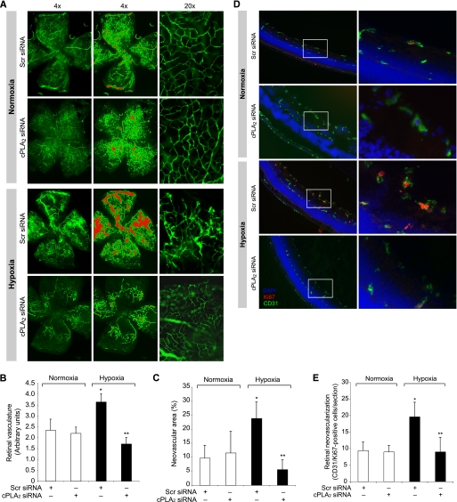
FIGURE 8.
Down-regulation of cPLA2 levels blocks hypoxia-induced retinal neovascularization. A, after exposure to 75% oxygen, the mice pups were returned to normoxia and administered 1 μg/0.5 μl scrambled (Scr) or cPLA2 siRNA at P13, P14, and P15 by intravitreal injections. At P17 the pups were anesthetized, perfused with FITC-dextran, sacrificed, and enucleated; retinas were isolated, and flat mounts of whole retinas were examined for retinal neovascularization (first column). Neovascular tufts are highlighted with a red color (second column). The third column shows the magnified sections of selected areas of the images shown in the first column. B and C, retinal vasculature was measured by fluorescence intensity in the total retinal area (B), and the neovascular area was calculated by the percentage of tufts area to total retinal area (C). D, all conditions are the same as in A except that cPLA2 siRNA was injected at P12 and P13, retinas were isolated at P15 and fixed, and cross-sections were made and analyzed by double immunofluorescence staining for Ki67 and CD31. The right column shows the higher magnification (×40) of the selected areas (enclosed in rectangles) of the images shown in the left column. E, retinal neovascularization was quantified by counting Ki67- and CD31-positive cells that extended anterior to the inner limiting membrane per section (n = 3 eyes, 5 sections/eye). The values are presented as mean ± S.D. *, p < 0.01 versus normoxia + scrambled (Scr) siRNA; **, p < 0.01 versus hypoxia + scrambled siRNA.

