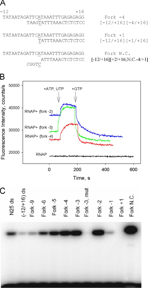
FIGURE 6.
Transcription activity of downstream fork junction probes. A, downstream fork junction probes used. B, shown is a beacon assay of transcription activity of [−12/+16][−2/+16], [−12/+16][−3/+16], and [−12/+16][−4/+16] downstream fork junctions. Time-dependent change of the fluorescence signal was measured upon the addition of ATP and UTP followed by the addition of GTP (final concentration of 0.5 mm each) to complexes of the fork junctions (2 nm) with 1 nm (211Cys-TMR) σ70 holoenzyme. C, shown is an abortive transcript synthesis by RNAP complexes with downstream fork junction probes. The Fork −3 mut template is a derivative of [−12/+16][−3/+16] fork junction with −11A and −7T substituted by non-consensus C; N25 ds and (−12/+16) ds represent double-stranded −60/+30 and −12/+16 fragments of the T5N25 promoter DNA.

