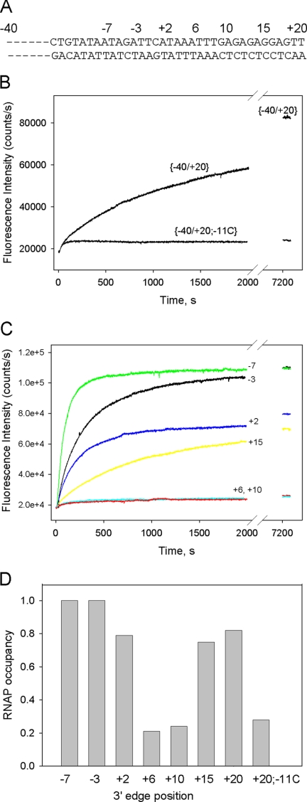
FIGURE 7.
Measuring RNAP interaction with double-stranded promoter fragments using protein beacon assay. A, shown is the primary sequence of a double-stranded [−40/+20] probe, based on the sequence of the T5N25 promoter. Other probes are derivatives of [−40/+20] truncated downstream at positions −7, −3, +2, +6, +10, or +15. B, shown is time dependence of the increase in fluorescence upon mixing 1 nm (211Cys-TMR) σ70 holoenzyme with 2 nm [−40/+20] and a derivative bearing a C to A substitution at position −11 [−40/+20;−11C]. C, time dependence of fluorescence upon mixing 1 nm (211Cys-TMR) σ70 holoenzyme with 2 nm [−40/−7] (green), [−40/−3] (black), [−40/+2] (blue), [−40/+6] (cyan), [−40/+10] (red), and [−40/+15] (yellow). D, RNAP occupancies measured in samples containing 1 nm RNAP beacon and 2 nm probes truncated at the indicated positions. The occupancies were determined using a competition binding assay described in the supplemental material.

