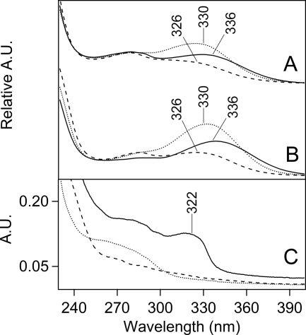
FIGURE 3.
Absorption spectra of peptides, derived from tryptic digestion of Tris-washed PSII. In A and B, the peptides were separated by HPLC. The 350-nm chromatogram exhibited three fractions with approximate retention times of 28 (fraction 1), 35 (fraction 2), and 36 (fraction 3) min (supplemental “Experimental Procedures” and Fig. S2). A, absorption spectra of fraction 1 (solid line), fraction 2 (dotted line), and fraction 3 (dashed line) from samples in which PSII was not treated with B5A. B, absorption spectra of fraction 1 (solid line), fraction 2 (dotted line), and fraction 3 (dashed line) from samples in which PSII was treated with B5A. Fraction 1 corresponds to a CP43 peptide. C, absorption spectra of two-dimensional gel-purified, B5A-labeled CP43 peptides before (dotted line) and after (solid line) affinity chromatography. The data in the dashed black line in C is the spectrum of B5A alone. Spectra shown in A and B were derived from the HPLC chromatogram and are on an arbitrary y scale. The spectra shown in C were measured on a Hitachi spectrophotometer. See “Experimental Procedures” for more information.

