FIG. 2.
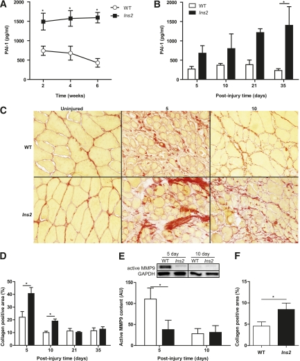
Type 1 diabetes causes elevated PAI-1, suppresses MMP9 activation, and increases collagen content during early regeneration time points. Significantly elevated PAI-1 levels in Ins2WT/C96Y mice compared with WT mice were found in blood plasma collected (A) throughout type 1 diabetes progression (main effect: P < 0.001; N = 16) and (B) after CTX injury (main effect: P < 0.001; N = 16). This led to the hypothesis that collagen would be elevated in the Ins2WT/C96Y mice because of suppression of the fibrinolytic pathway. C: Picrosirius red staining of injured TA sections revealed increased collagen (red color), which was statistically significant (D, main effect: P = 0.001, interaction: P = 0.004; N = 16). E: MMP9, an important protease in skeletal muscle collagen cleavage, was also found to be significantly repressed in Ins2WT/C96Y mice (labeled Ins2, E) at the 5-day time point (N = 8). F: The short-term Ins2WT/C96Y mice also exhibited increased collagen at 10 days postinjury (t test: P = 0.047, N = 3). *Differences between groups at specific time points identified by Bonferroni post hoc analysis after 2-way ANOVA (A, B, D, E). A–E: White bars/circles represent WT, and black bars/squares represent Ins2WT/C96Y. (A high-quality digital representation of this figure is available in the online issue.)

