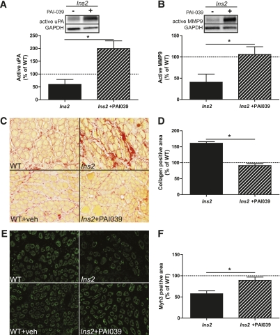
FIG. 3.
Pharmacologic treatment against PAI-1 improves fibrinolytic pathway activity, collagen degradation, and regeneration at 5 days post-CTX injury in type 1 diabetes. A: Treatment with PAI-039 caused an increase in free uPA in Ins2WT/C96Y compared with untreated Ins2WT/C96Y (t test: P = 0.004; N = 4). B: Similarly, active MMP9 was elevated in PAI-039–treated Ins2WT/C96Y (t test: P = 0.025; N = 4). These findings are characteristic of restored fibrinolytic pathway activity, which presumably led to the (C and D) reduced collagen levels (t test: P < 0.001; N = 4) and (E and F) increased Myh3-positive area (t test: P = 0.011; N = 4) observed in PAI-039–treated Ins2WT/C96Y compared with untreated Ins2WT/C96Y (labeled Ins2, C and E). A–E: Black bars represent Ins2WT/C96Y, and striped bars represent PAI-039–treated Ins2WT/C96Y. Data are presented relative to the mean of the WT and WT + vehicle pooled data. *Differences between groups identified by t test. (A high-quality digital representation of this figure is available in the online issue.)

