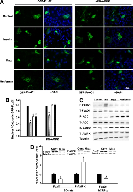
FIG. 4.
Nuclear exclusion and decrease in cellular FoxO1 induced by Mαα. A and B: HepG2 cells grown on cover slips were transfected with expression plasmids for GFP-FoxO1(TSS) in the absence or presence of DN-AMPK as indicated. After transfection (24 h), the culture medium was changed to serum-free DMEM supplemented with vehicle (black bars), 0.1 μmol/L insulin (hatched bars), 200 μmol/L Mαα (empty bars), or 2.0 mmol/L metformin (cross-hatched bars) as indicated, and further incubated for 24 h before fixation and DAPI staining. A: Representative immunofluorescence slides of GFP-FoxO1 (green) and DAPI-stained nuclei (blue; bar 20 μm). B: Nuclear-to-cytosolic ratio analyzed using fluorescence microscopy (Axioskop). Mean ± SE of three independent experiments. *Significant as compared with vehicle-treated cells (P < 0.05). C: HepG2 cells were incubated for 3 h in serum-free DMEM supplemented with 0.1 μmol/L insulin, 200 μmol/L Mαα, or 2.0 mmol/L metformin as indicated. Cell extracts were subjected to SDS-PAGE followed by Western blotting as described in research design and methods. Blots were probed with anti- P-FoxO1(T24), FoxO1, P-AMPK(T172), AMPK, P-ACC(S79), ACC, or tubulin antibodies. Representative blots of three independent experiments. D: SD rats were dosed daily by gavage for 2 weeks with vehicle (black bars) or with 80 mg Mαα/kg BW in 1% CMC (empty bars). hCRP transgenic mice were dosed with 0.06% (W/W) Mαα mixed in the diet for a period of 3 weeks (empty bars) or kept untreated (black bars). Liver extracts were subjected to SDS-PAGE followed by Western blotting as described in research design and methods. Blots were probed with anti-FoxO1 antibodies normalized to tubulin or P-AMPK(T172) antibodies normalized to AMPK. Hepatic FoxO1 content normalized to tubulin and P-AMPK(T172)-to-AMPK ratio of nontreated animals is defined as 1.0. Mean ± SE (n = 4 to 5). *Significant as compared with nontreated animals (P < 0.05). Inset: Representative blots. (A high-quality digital representation of this figure is available in the online issue.)

