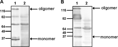
FIGURE 2.
Expression of nadA in Y. enterocolitica. Western blot analysis of outer membrane fractions (10 μg) incubated at 100 °C. A, WA-c Δinv(pnadA) (lane 1); WA-c Δinv(p) (lane 2). B, WA-314 Δyad:nadA (lane 1); WA-314 ΔyadA (lane 2). The assays were performed using rabbit anti-NadA serum and secondary peroxidase-conjugated antibody. The arrows indicate the oligomeric and monomeric form of NadA.

