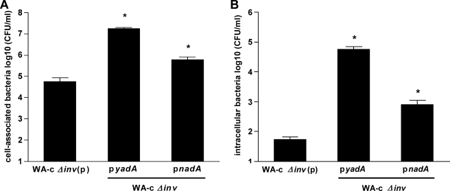
FIGURE 4.
Role of nadA-expressing yersiniae in adhesion and invasion into Chang cells. Chang cell monolayers were infected with WA-c Δinv(p) (negative control), WA-c Δinv(pyadA), and WA-c Δinv(pnadA) for 3 h (moi 100). Shown are (A) total cell-associated bacteria (including both intra- and extracellular bacteria), and (B) intracellular bacteria determined using gentamicin protection assays. The number of cell-associated and intracellular bacteria is expressed as log10 colony forming units per ml (CFU/ml). Data are expressed as the means ± S.E. of the mean of at least three independent experiments. *, p < 0.0383 versus WA-c Δinv(p).

