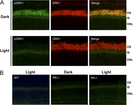
FIGURE 3.
Immunocytochemical analysis of phosphorylated GRK1 in dark- and light-adapted mice. Mice were dark- or light-adapted as described in the legend for Fig. 2, followed by euthanasia and removal of the eyes. Eyecups were fixed as described under “Materials and Methods. ” A, sections from dark- and light-adapted wild-type (WT) mouse retinas were double-labeled with anti-pGRK1 and anti-GRK1. B, retina sections from light-adapted WT mice and dark-reared GRK1 knock-out (RK−/−) mice, which were euthanized and enucleated in the dark or light-adapted prior to euthanasia, were labeled with anti-pGRK1. OS, outer segment; IS; inner segment; ONL; outer nuclear layer.

