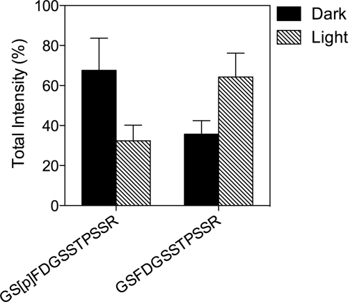
FIGURE 4.
Mass spectrometry analysis of GRK1 phosphorylated on Ser21 in photoreceptor cells. Retinas from dark- and light-adapted rats were processed for serial, tangential sectioning as described under “Materials and Methods. ” The sections were compared by mass spectrometry for changes in levels of a phosphopeptide corresponding to amino acids 20–31 of rat GRK1 phosphorylated on Ser21 compared with the corresponding unphosphorylated peptide. Error bars represent the range of duplicate dark and light analyses.

