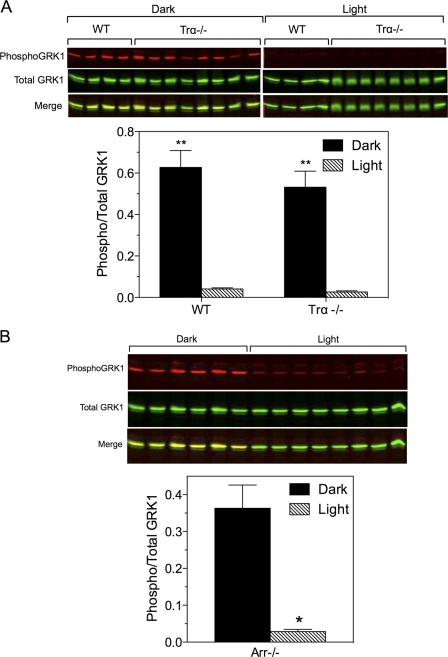
FIGURE 8.
Phosphorylation of GRK1 and phosducin in Trα−/− and Arr−/− mice. A, wild-type and Trα−/− mice were dark-adapted overnight, then maintained in the dark or exposed to light for 2–3 h, euthanized, and the retinas were dissected for immunoblot analysis. Upper panel, Western blot of samples collected from two experiments. Lower panel, quantitative analysis of data shown above. Error bars represent S.E., n = 4 for each WT group; n = 8 for each Trα−/− group; **, p < 0.001 dark versus light. B, Arr−/− mice were dark-adapted overnight and exposed to light for 30–40 min. Upper panel, Western blot of samples collected from a single experiment. Lower panel, quantitative analysis of the data shown above. n = 6 (dark) and 8 (light); *, p < 0.0001 versus dark.

