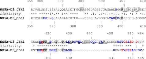
FIGURE 1.
Sequence comparison of NS5A-D3 from JFH-1 and Con1 HCV strains and interaction with cyclophilin A. The NS5A-D3 sequence from the HCV JFH-1 strain (aa 355–464; genotype 2a; GenBankTM accession number AB047639) is aligned with the NS5A-D3 sequence from the HCV Con1 strain (aa 359–445; genotype 1b; AC number, AJ238799). Amino acids are numbered with respect to the respective NS5A proteins. The hyphens indicate gaps compared with the sequence alignment. Similarities between NS5A sequences are depicted according to ClustalW convention (asterisk, invariant; colon, highly similar; dot, similar). Proline residues (which are not visible in the 1H-15N HSQC NMR experiment; see “Experimental Procedures”) are highlighted in gray, and those conserved in both strains are in boldface type. The perturbations of chemical shifts observed for each residue in the 1H,15N HSQC spectrum of [15N]NS5A-D3 (100 μm) in the presence of cyclophilin A (100 μm) are indicated as follows: residue in blue, chemical shift perturbation and/or decreased intensity; residue in red, broadening of the resonance beyond detection; residue underlined, residue with its minor form perturbed (chemical shift perturbation and/or decreased intensity).

