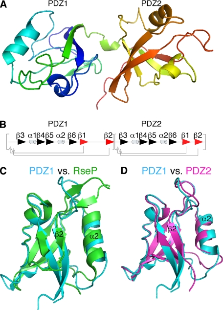
FIGURE 1.
Structure of the GRASP domain and comparison of its PDZ domains. A, graphic structure of GRASP55 1–208 showing globular domains of PDZ1 (left) and PDZ2 (right) separated by a short α-helix. B, ordering of secondary structural elements within GRASP55 PDZ1 and PDZ2 indicates a circular permutation of a typical eukaryotic PDZ domain. C, graphic representation of GRAS55 PDZ1 (blue) and RseP PDZ2 (green) demonstrates high structural overlap. D, comparison of G55 PDZ1 (blue) and G55PDZ2 (purple) shows high overall similarity with one another.

