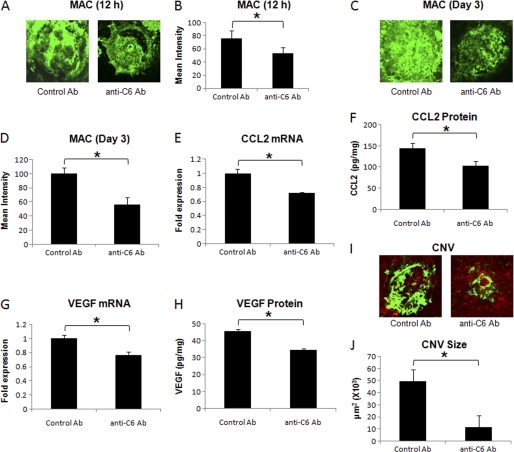
FIGURE 2.
Effect of i.p. injection of anti-murine C6 on MAC deposition, CCL2 expression, VEGF expression, and formation of CNV complex. Representative confocal micrographs of RPE-choroid-sclera flat mounts immunostained for MAC at 12 h (A) and day 3 (C) post-laser treatment from mice injected i.p. with anti-murine C6 or control antibody. Graphs show semi-quantitative evaluation of positive fluorescent signal for MAC at 12 h (B) and at day 3 (D). CCL2 mRNA (E) and protein (F) levels were analyzed 12 h after laser treatment, whereas levels of VEGF mRNA (G) and protein (H) were determined at day 3 post-laser treatment by real-time RT-PCR and ELISA, respectively, in RPE-choroid of mice treated i.p. with anti-murine C6 or control antibody. Representative confocal microphotographs of RPE-choroid-sclera flat mounts with FITC-dextran perfused vessels (green) from C57BL/6 mice injected i.p. with anti-murine C6 or control antibody and sacrificed at day 7 post-laser treatment are shown (I). Cumulative data obtained by the quantification of the images are shown in J. Quantification and statistical analyses, including S.D. and Student's t test, were performed as described under “Experimental Procedures.” All data are representative of three independent experiments. *, p < 0.05.

