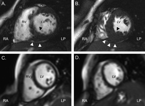
FIGURE 3.
Cardiac magnetic resonance findings. All frames were taken at end-diastole. A, short-axis cut at mid-ventricular level is shown. B, short-axis cut at a more apical level is shown. C and D, short axis cuts at comparable levels to A and B in a sex- and age-matched subject with normal cardiac anatomy and function are shown. Note the fine structure of the left ventricular myocardium. RA, right anterior; LP, left posterior; RV, right ventricle; LV, left ventricle. Arrowheads denote focal thickening of an inferior septal segment.

