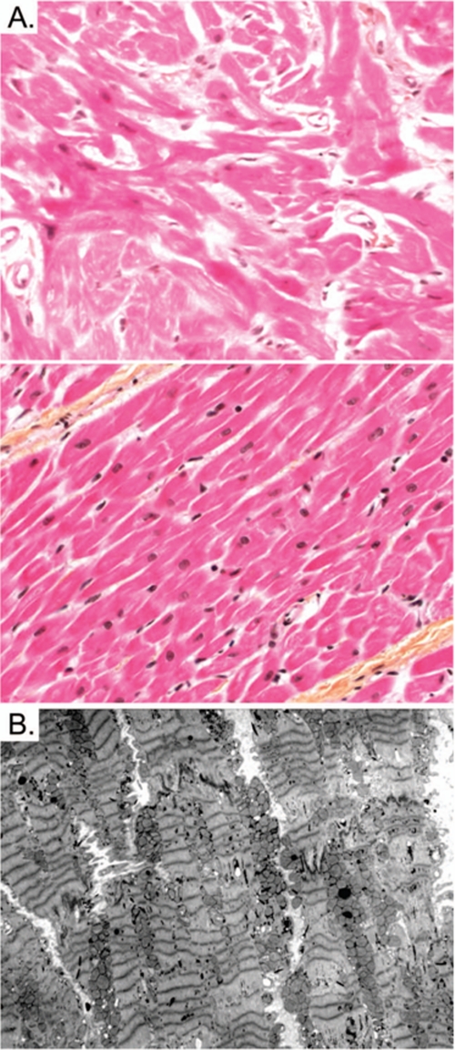
FIGURE 5.
Histopathological studies. A, extensive cardiomyocyte disarray is seen in light microscopy using hematoxylin phloxine saffron stain (top) in comparison with normal histology (bottom) at 400×-fold magnification. B, transmission electron microscopy shows extensive Z-disc streaming but normal mitochondrial morphology.

