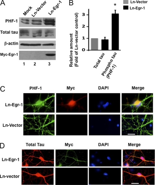
FIGURE 9.
Overexpression of Egr-1 in rat hippocampal primary neurons in culture promotes tau phosphorylation but does not change tau distribution. Rat primary neurons in culture were infected with the indicated virus and then analyzed by either Western blot analysis or immunocytochemistry. A, Western blots. B, relative amounts. The relative amount of phosphorylated tau was determined as in Fig. 2 using a Western blot representing A. Values are the mean ± S.E. from three independent cultures. *, p > 0.05 (t test). C and D, immunocytochemistry. Shown are infected neurons stained for PHF-1 (phosphorylated tau), Myc (Myc-Egr-1), DAPI (nucleus), Tau 5 (total tau), and merge (co-localization). Scale bars, 15 μm.

