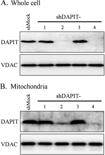
FIGURE 1.
Expression of DAPIT protein in whole cells (A) and the isolated mitochondria of the mock-treated (shMock) and DAPIT-knockdown (shDAPIT-1, 2, 3, 4) HeLa cells (B). Whole cell (20 μg of protein) and mitochondria (5 μg of protein) were analyzed with SDS-PAGE, and DAPIT was stained with Western blotting. VDAC was also stained as an internal standard of mitochondrial protein. Experiments were carried out at least four times, and representative results are shown.

