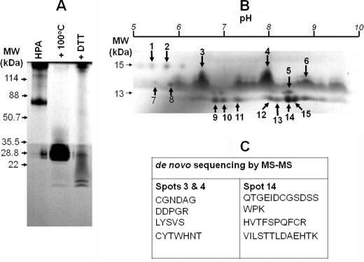
FIGURE 1.
Analysis of HPA lectin purified from the albumen gland of the Roman snail, H. pomatia, highlighted the complexity of the preparation. A, separation using Tris-Tricine SDS-PAGE at 180 V for 35 min. The lanes indicate native HPA, HPA after denaturation for 5 min at 100 °C, and HPA after treatment with 0.2 m DTT for 15 min at 20 °C. 25 μg of protein was loaded into each well. The protein was visualized by staining the gel with Coomassie Brilliant Blue. B, two-dimensional separation of 150 μg of HPA preparation using a 3–10 pH linear immobilized pH gradient strip and SDS-PAGE as for A. The spots indicated by the arrows and identified by Coomassie Brilliant Blue staining were used for MS/MS analysis. C, amino acid sequence data for spots 3, 4, and 14 as indicated.

