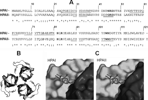
FIGURE 2.
Pairwise sequence alignment and structure prediction of HPAI and HPAII. A, pairwise alignment of the amino acid sequences for HPAI and HPAII using ClustalW. Identical amino acids are indicated with an asterisk, amino acids that form the carbohydrate-binding site are shown in bold, and amino acids that form hydrogen bonds during trimer formation are shown in italics. The amino acid residues identified by MS/MS are underlined. B, ribbon representations of HPAI (blue) and HPAII (green) viewed from the top. GalNAc is shown in red. C, surface representation of the HPAI and HPAII GalNAc binding site. The binding pocket is formed at the interface of the two monomeric domains, shown in blue and green. GalNAc is shown in red.

