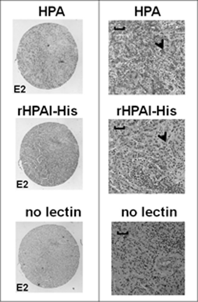
FIGURE 6.

Light micrographs of serial sections of an infiltrating breast carcinoma tissue stained with native and rHPAI-His and counterstained with hematoxylin. The sections were incubated with either biotinylated native HPA, biotinylated rHPAI-His, or processed without addition of lectin, as indicated. The left panel shows the low magnification view (×4 objective), and the right panel shows a higher magnification view (×20 objective). Scale bars, 100 μm. Lectin binding was visualized following incubation with streptavidin-peroxidase by the addition of the chromogenic substrate diaminobenzidine to give a brown-colored precipitate (shown by the arrowheads). Nuclei were stained with Harris hematoxylin.
