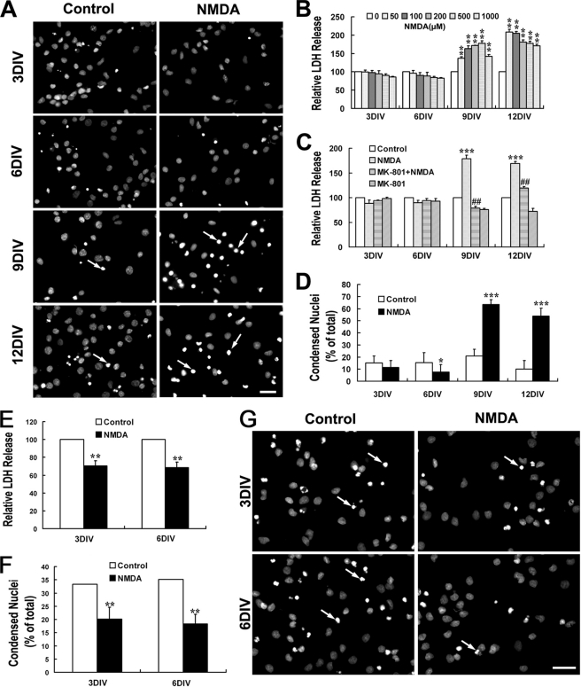
FIGURE 1.
NMDA triggers dramatic neurotoxicity in mature hippocampal neurons although it is neuroprotective for immature neuron. At different DIV, cultured hippocampal neurons were treated with or without NMDA (100 μm) for 15 min in modified Locke's solution and washed three times with DMEM and then incubated with the original culture medium. LDH release measurements and Hoechst staining were carried out 24 h later. NMDA neurotoxicity increased significantly during neuron maturation (A–D). A, representative images of Hoechst staining showing the condensed nuclei (arrow) after treatment with NMDA (100 μm) or not. B, NMDA-induced neuronal release of LDH. **, p < 0.01 versus control (0). C and D, effect of MK-801 on NMDA-induced LDH release (C) and nuclei condensation (D). *, p < 0.05; ***, p < 0.001 versus control; ##, p < 0.01 versus NMDA. NMDA is neuroprotective against tropic deprivation in 3- and 6DIV immature neurons (E–G). Neurons were incubated in DMEM without any nutrition supplement after NMDA challenge or not (DMEM protocol). Considerable cell death could be seen in the control group. E and F, NMDA-induced neuronal of LDH release (E) and nuclei condensation (F). **, p < 0.05 versus control. G, representative images of Hoechst staining showing the condensed nuclei (arrow) in groups treated with NMDA (100 μm) or not. Bar, 20 μm.

