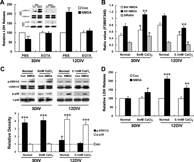
FIGURE 10.
NMDA neurotoxicity but not NMDA induced ERK1/2 and p38 MAPK activation changes according to extracellular calcium level. For LDH release assay, 3- and 12DIV neurons were treated with or without NMDA (100 μm) for 15 min in normal or calcium-free EGTA (5 mm) containing low (0.1 mm) and high (5 mm) calcium (A) and Locke's solution (D) and washed three times with DMEM and then incubated with original culture medium (A, 3DIV) or DMEM for 24 h before LDH release assay. For MAPK activation, 3- and 12DIV neurons were starved in DMEM for 6 h and treated with or without NMDA (100 μm) for 15 min in normal or calcium-free EGTA (5 mm) containing low (0.1 mm) and high (5 mm) calcium (A) and Locke's solution (C). Immunoblotting was done by using anti-phosphorylated p38 or ERK1/2 antibodies (p-p38, t-ERK1/2) and anti-total p38 or ERK1/2 (t-p38, p-ERK1/2) antibodies, with the latter serving as an internal control for protein loading to assess the activation level of p38 and ERK1/2. For calcium imaging, 3- and 12DIV neurons were perfused with low (0.1 mm) or high (5 mm) calcium BSS for 2 min and then with the same BSS containing NMDA (100 μm) or not for 3–5 min. A, both NMDA induced neuroprotection and neurotoxicity as well as ERK1/2 and p38 activations are calcium-dependent. **, p < 0.05 versus Control (Con). Note that EGTA treatment abolishes both NMDA-induced neuroprotection and ERK1/2 activation in 3DIV neurons, as well neurotoxicity and p38 activation in 12DIV neurons. B, basal [Ca2+]i in 3- and 12DIV neurons can be adjusted to a similar level by changing the extracellular concentration of CaCl2. *, p < 0.05; **, p < 0.01 versus normal. Note the elevated basal [Ca2+]i level and enhanced calcium influx in 3DIV neuron under 5 mm CaCl2 condition, as well as the descended basal [Ca2+]i level and decreased calcium influx in 12DIV neuron under 0.1 mm CaCl2 conditions, which have no significant differences with each other. There are no significant differences in [Ca2+]i levels between 3DIV neurons under normal conditions and 12DIV neurons under 0.1 mm CaCl2, as well as between 12DIV neurons under normal and 3DIV neuron under 5 mm CaCl2 condition both before and after NMDA stimulation. C, pattern of NMDA-induced activation of ERK1/2 and p38 in mature and immature neurons did not change with extracellular calcium level. Representative blots are shown from three independent experiments. *, p < 0.05; ***, p < 0.001 versus control. Note the dramatic activation of ERK1/2 but not p38 in 3DIV neurons under both normal and high extracellular calcium (5 mm) conditions, as well as the strong activation of p38 but not ERK1/2 in 12DIV neuron under both normal and low extracellular calcium (0.1 mm) conditions. D, neurotoxicity is affected by extracellular calcium levels. *, p < 0.05; ***, p < 0.001 versus control; **, p < 0.01 versus normal. Note that NMDA triggers significant LDH release in 3DIV neurons under 5 mm CaCl2 conditions but not normal conditions, and LDH release in 12DIV neuron is significantly decreased under 0.1 mm CaCl2 conditions.

