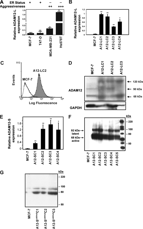
FIGURE 2.
ADAM12 expression is elevated in aggressive breast cancer cells. A, real-time RT-PCR analysis indicated significantly higher expression of ADAM12-L in highly aggressive ER-negative cells as compared with ER-positive, less invasive breast cancer cells. *, p = 0.005; **, p ≤ 0.0001. B–G, stable expression of ADAM12 isoforms in MCF-7 cells. Individual ADAM12-l-expressing clones are indicated as LC1, LC2, LC3, and LC4. B, real-time RT-PCR analysis of ADAM12-L in WT MCF-7 and representative ADAM12-L-overexpressing clones. *, p = 0.001; **, p ≤ 0.0001. Cell-surface ADAM12-L expression was confirmed via FACS analysis. C, a representative ADAM12-L clone, LC2, had considerably higher cell surface staining as compared with the WT MCF-7. D, immunoblot analysis of cell lysates indicating increased expression of ADAM12-L in transfectants as compared with WT MCF-7 cells. Three distinct bands representing ADAM12-L were detected, including an ∼120-kDa latent form, an ∼90-kDa active form, and an ∼68-kDa truncated form, respectively. E, ADAM12-S stable clones displayed an ∼10- to 30-fold increase in mRNA expression as compared with WT MCF-7 cells. *, p ≤ 0.01; **, p ≤ 0.0001. Immunoblot of serum-free CM demonstrated expression of latent and the active species of ADAM12-S (F) and ADAM12-Scatmut (G) in the clones but not WT MCF-7 cells.

