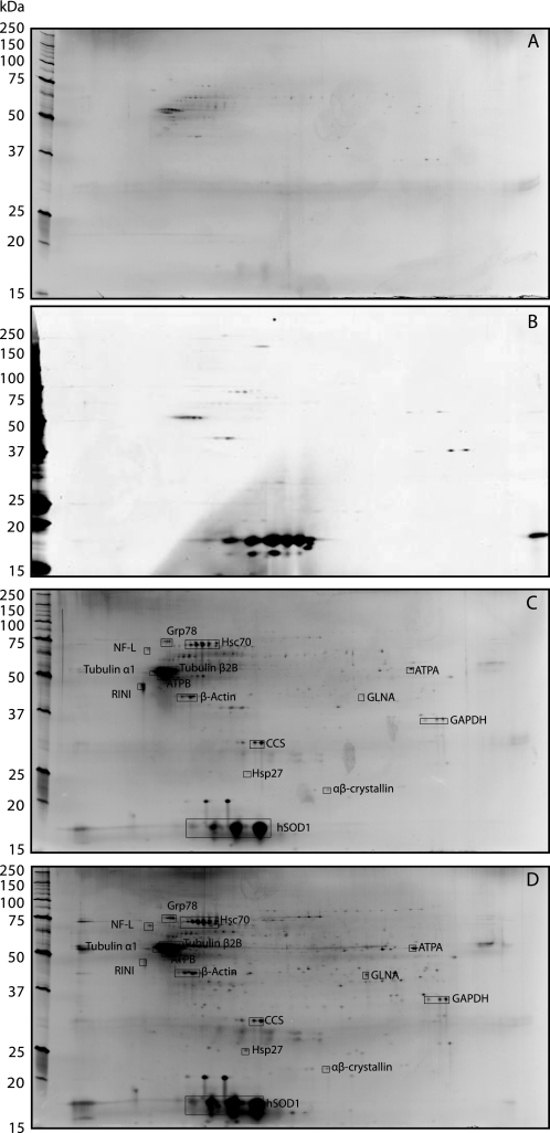
FIGURE 3.
Two-dimensional gel patterns of rat spinal cord extract proteins binding to immobilized holo- and apo-hSOD1. A, proteins bound to ethanolamine-blocked, B, wild-type holo-hSOD1-bound C, wild-type apo-hSOD1-bound, and D, G93A apo-hSOD1-bound Sepharose gels (see “Experimental Procedures”) were visualized by two-dimensional electrophoresis and silver staining. Proteins identified with MALDI-TOF/MS are shown. In A, C, and D, 10–14.5% gels were used for the second dimension and a 12.5% gel in B.

