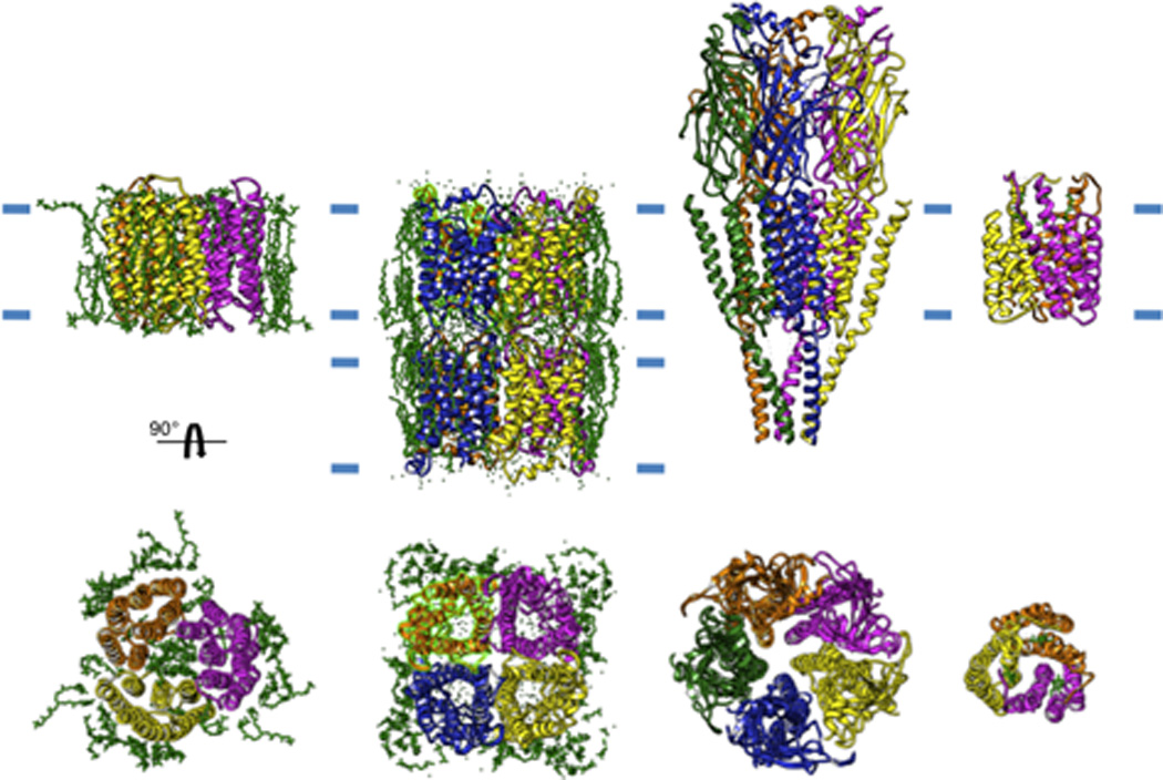
Figure 2. Selection of high-resolution membrane protein structures solved by electron crystallography.

From left to right: a trimer of bacteriorhodopsin, a double-layer of the tetrameric AQP0, the heteropentameric acetylcholine receptor and a trimer of gluthathione transferase 1. The approximate boundaries of the bilayer is indicated by short blue lines and individual lipid molecules present in the structure are shown in green.
