
Figure 4. Pipeline for protein structure determination by electron crystallography.
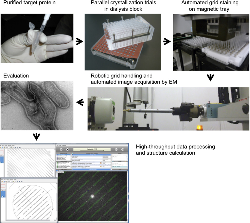
Target membrane proteins are purified in detergent micelles in a stable and monodisperse form. Following the addition of lipids to form mixed micelles of protein, detergent and lipid, the crystallization process is studied by removing dialysis in a 96-well dialysis block. The 96 crystallization conditions are harvested, transferred to EM grids and negative stained with a liquid-handling robot. The EM grids are robotically inserted into the electron microscope, and images are recorded automatically and stored in a database. Thus, a broad range of parameters can be explored in an attempt to find large, well ordered crystals. Finally, image processing and structure determination can be carried out using, for example, the 2dx software.