
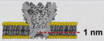
Table 2.
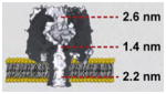
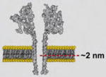
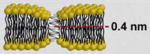
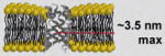
Selection of commonly applied biological pores in nanobiotechnology. Note, all illustrations of pores and lipids are drawn to scale to facilitate the comparison of their sizes
| Pore | Source | Pore assembly | La | Illustration | ø | |
|---|---|---|---|---|---|---|
| Large Proteins | α-hemolysin [69–71] | Staphylococcus aureus bacterium | Heptameric Pore |
|
||
| aerolysin [72] | Aeromonas hydrophila bacterium | Heptameric Pore |
|
|||
| anthrax toxin [73] | Bacillus anthracis bacterium | Heptameric Pore |
|
|||
| diphtheria toxin [4•] | Corynebacterium diphtherian bacterium | Monomeric | not shown | |||
|
| ||||||
| Small Peptides | gramicidin A [33•,74–76] | Bacillus brevis bacterium | Head-to-head dimerization |
|
||
| alamethicin [77,78] | Trichoderma viride fungus | Bundle of α-helices (4–11) |
|
|||
| melittin [79,80] | Apis mellifera bee venom | Bundle of α-helices | similar to alamethicin | |||
|
| ||||||
| Porins | MspA [30•] | Myobacterium smegmatis bacterium | Octameric Pore |
|
||
| OmpG [81] | Escherichia coli bacterium | Monomeric Pore |

|
|||
a
Length of the constriction zone within the lumen of the pore.