
Table 3.
Commonly applied model lipid membranes for reconstitution and application of biological pores in nanobiotechnology
| Platform | Description | Illustration | Typical Application |
|---|---|---|---|
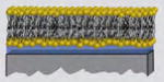
| Supported Lipid Bilayer | Bilayer supported on a solid substrate |
|
Incorporation of biological pores changes the electrical impedance of the supported bilayers. Binding to these pores can be detected by additional changes in impedance. |
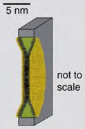
| Planar Lipid Bilayer | Bilayer spanning a small pore between two aqueous solutions |
|
Reconstitution of ion channel proteins or pore-forming peptides changes the ionic conductance across the bilayer. Ion currents through individual pores as well as changes in conductance due to the presence of analytes can be detected. |
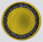
| Liposomes | Lipid bilayer vesicles suspended in aqueous solutions |
|
Reconstitution of biological pores permeabilizes liposomal membranes; cargo molecules encapsulated inside the liposomes can be released through these pores. |
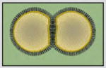
| Droplet Interface Bilayer | Lipid bilayer formed at the interface of two aqueous droplets that are coated with a monolayer of lipids within an oil phase |
|
Similar to planar lipid bilayers. Incorporation of biological pores changes the ionic conductance across the bilayer. Ionic currents through individual pores can be detected by inserting an electrode into each droplet. |