Fig. 3.
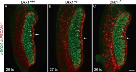
Testis development in the Dkk1-null embryos appears normal morphologically. A Immunofluorescent analysis of whole-mount UGRs from wild-type (Dkk1+/+), B Dkk1+/–, and C Dkk1–/– embryos (12.5 dpc). SOX9 (green) marks Sertoli cells, PECAM (red) marks germ cells and vasculature. No delay or disruption of testis cord formation is seen (asterisks). Endothelial cells localise to the appropriate area in Dkk1-null gonads and display a similar level of organisation to staged-matched controls (arrows). Tail somite (ts) stages for each sample are indicated. Scale bar = 100 μm.

