Figure 2.
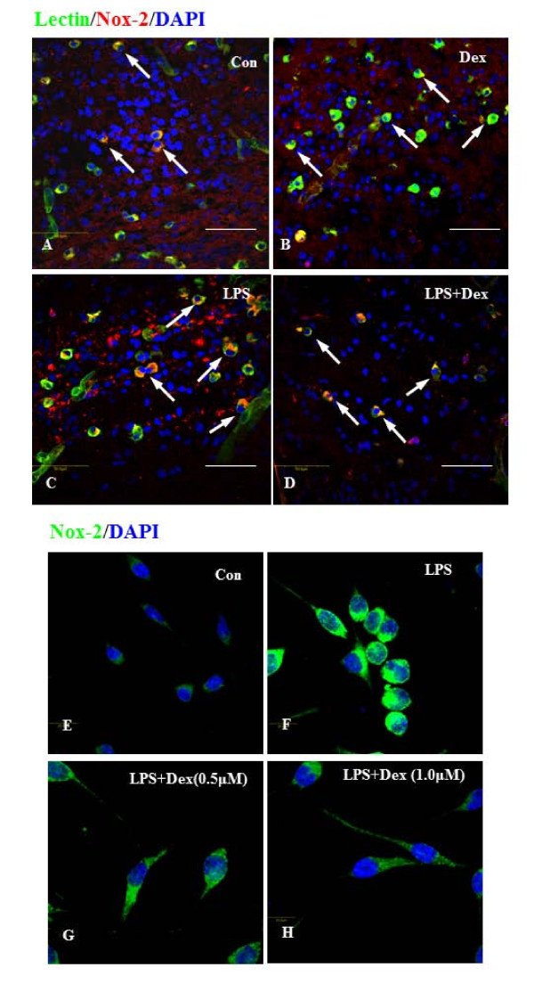
A-H. Confocal images showing the suppressive effect of Dex on Nox-2 expression in LPS-treated microglia in vivo and in vitro. Colocalization of lectin (green), Nox-2 (red) and DAPI (blue) was detected in corpus callosum region of brain sections from 3d postnatal rats with or without LPS or LPS+Dex injection (A-D). The incidence of Nox-2-positive microglial cells (arrows) was increased in the brain of LPS-injected rats (C) compared to control (A) and to Dex alone (B)and the frequency of Nox-2-positive cells substantially decreased after Dex treatment (D). Double labeling was also carried out between Nox-2 (green) and DAPI (blue) in BV-2 cells treated with LPS for 6 h without (F) or with Dex (G, 0.5 &H, 1 μM) in vitro. The Nox-2 immunoreactivity appears to be increased in cells treated with LPS (F), compared to control (E). However, Dex decreased the intensity of immunoreactivity in a dose dependent manner in cells treated with LPS (G, H).

