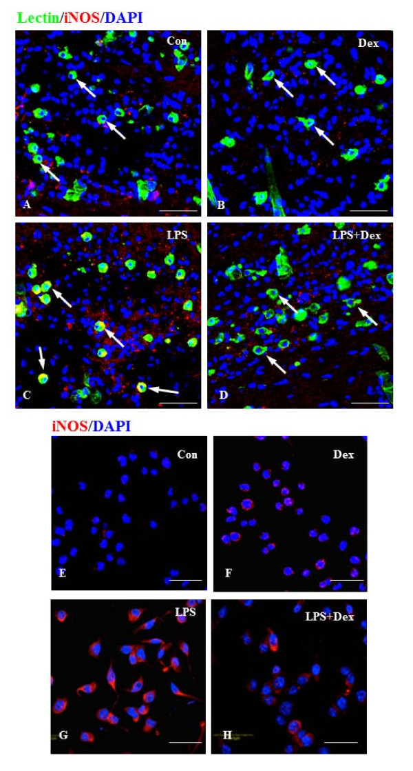
Figure 4.
A-H. Suppressive effect of Dex on iNOS expression in LPS-activated microglia in the corpus callosum of 3 day old rat brain and the BV-2 cells in vitro. Immunofluorescence analysis shows that iNOS (red) expression, which was hardly detectable in control cells (A, E) and cells treated with Dex alone (B, F). The induction of iNOS immunoexpression was evident in cells exposed to LPS (C, G). The induction was suppressed upon treatment with Dex (D, H). Note that the cells were counterstained with DAPI (blue). Scale bar (A-H); 50 μm

