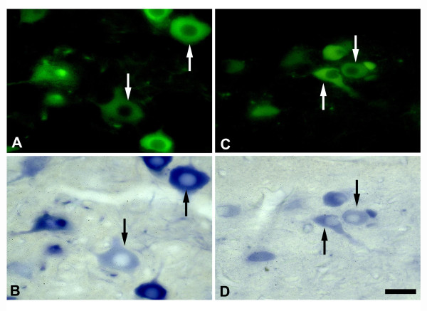
Figure 1.
NADPH-d/nNOS colocalization studies. Fluorescence (A, C) and light (B, D) photomicrographs showing nNOS(+) (A, C) and NADPH-d(+) (B, D) labeled neurons on the lesioned side of the HN (A, B) and DMN (C, D) of rats following crush injury of left vagus and hypoglossal nerves. Note that a most of nNOS(+) neurons (arrows in A-D) contain NADPH-d in the HN and DMN. Scale bar = 50 μm.

