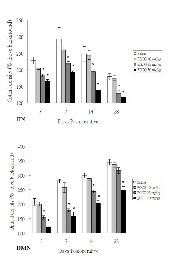
Figure 5.
Mean optical density (OD) of NADPH-d(+) neurons in the HN and DMN of adult rats at 3, 7, 14 and 28 days after PNCI and EGCG treatment. The changes of mean optical density (determining staining intensity) in NADPH-d(+) neurons of examined nuclei show a similar trend to the population modification of labeled neurons. The OD value in the HN of high dose EGCG-treated (25 and 50 mg/kg) animals is much lower than those of low dose EGCG-treated (10 mg/kg) or non-treated ones at 7, 14 and 28 days after PNCI. The EGCG-mediated noticeable decrease of NADPH-d optical density is also evidenced in the DMN at different time points after PNCI. *, p < 0.05 for the comparison between EGCG-treated and saline-treated groups at the same survival time point.

